Anti-pan Cytokeratin antibody PCK-26 鼠单克隆[PCK-26
- 产品型号: abcam ab6401
- 简单描述
- Anti-pan Cytokeratin antibody [PCK-26] 鼠单克隆[PCK-26DescriptionMouse monoclonal [PCK-26] to pan CytokeratinTested applicationsWB, IHC-FoFr, IHC-P, IHC-Fr, IF, Dot Blot, Flow Cyt, ICC/IFCro
详细介绍
Anti-pan Cytokeratin antibody [PCK-26] 鼠单克隆[PCK-26
Form
Liquid
Storage instructions
Store at +4°C short term (1-2 weeks). Aliquot and store at -20°C or -80°C. Avoid repeated freeze / thaw cycles.
Storage buffer
Preservative: 15mM Sodium Azide
Constituents: Ascites
Concentration
Anti-pan Cytokeratin antibody [PCK-26] 鼠单克隆[PCK-26
Purity
Ascites
Clonality
Monoclonal
Clone number
PCK-26
Myeloma
unknown
Isotype
IgG1
Light chain type
unknown
Research areas
Signal Transduction >> Cytoskeleton / ECM >> Cytoskeleton >> Intermediate Filaments >> Class I >> Cytokeratins
-
Immunohistochemistry (Formalin/PFA-fixed paraffin-embedded sections) – pan Cytokeratin antibody [PCK-26] (ab6401)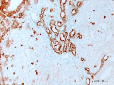 (enlarge)
-
Immunohistochemistry (Formalin/PFA-fixed paraffin-embedded sections) – pan Cytokeratin antibody [PCK-26] (ab6401) (enlarge)
-
Western blot – pan Cytokeratin antibody [PCK-26] (ab6401) (enlarge)
Applications
Show applications key
Our Abpromise guarantee covers the use of ab6401 in the following tested applications.
The application notes include recommended starting dilutions; optimal dilutions/concentrations should be determined by the end user.
- ShowHide4 Images
WB
Four stars(5 Abreviews)WB: Use at an assay depe…Read more →
WB: Use at an assay dependent dilution.
- ShowHide
IHC-FoFr
IHC-FoFr: Use at an assa…Read more →
IHC-FoFr: Use at an assay dependent dilution.
- ShowHide3 Images
IHC-P
Five stars(3 Abreviews)IHC-P: Use at an assay d…Read more →
IHC-P: Use at an assay dependent dilution.
- ShowHide2 Images
IHC-Fr
Four stars(3 Abreviews)IHC-Fr: Use at an assay …Read more →
IHC-Fr: Use at an assay dependent dilution.
-
IF
IF: 1/300.
IF: 1/300.
- ShowHide
Dot Blot
Dot: Use at an assay dep…Read more →
Dot: Use at an assay dependent dilution.
- ShowHide
Flow Cyt
See more…Read more →
Flow Cyt: Use at an assay dependent dilution. PubMed: 19855980Use 10µl for 5 x 10⁷ cells.
- 2 Images
ICC/IF
Five stars(2 Abreviews)ICC/IF: 1/300.
ICC/IF: 1/300.
Target
Relevance
Cytokeratins, a group comprising at least 29 different proteins, are characteristic of epithelial and trichocytic cells. Cytokeratins 1, 4, 5, 6, and 8 are members of the type II neutral to basic subfamily. Monoclonal anti cytokeratins are specific markers of epithelial cell differentiation and have been widely used as tools in tumor identification and classification. Monoclonal Anti Pan Cytokeratin (mixture) is a broadly reactive reagent, which recognizes epitopes present in most human epithelial tissues. It facilitates typing of normal, metaplastic and neoplastic cells. Synergy between the various components results in staining amplification. This enables identification of cells, which would otherwise be stained only marginally. The mixture may aid in the discrimination of carcinomas and nonepithelial tumors such as sarcomas, lymphomas and neural tumors. It is also useful in detecting micrometastases in lymph nodes, bone marrow and other tissues and for determining the origin of poorly differentiated tumors. There are two types of cytokeratins the acidic type I cytokeratins and the basic or neutral type II cytokeratins. Cytokeratins are usually found in pairs comprising a type I cytokeratin and a type II cytokeratin. Usually the type II cytokeratins are 8kD larger than their type I counterparts.
Cellular localization
Cytoplasmic
Anti-pan Cytokeratin antibody [PCK-26] images:
Immunohistochemistry (Formalin/PFA-fixed paraffin-embedded sections) – pan Cytokeratin antibody [PCK-26] (ab6401)

ab6401 staining pan Cytokeratin in Mouse liver tumor tissue sections by IHC-P (Paraformaldehyde fixed paraffin embedded sections). Tissue was fixed with paraformaldehyde and blocked with maleate buffer blocking solution for 30 minutes and 22°C. Antigen retrieval was by heat mediation in citrate buffer. Samples were incubated with primary antibody (1/500) in maleate buffer blocking solution for 16 hours at 22°C. An undiluted biotin-conjugated Donkey polyclonal to mouse IgG was used as secondary antibody.
This image is courtesy of an Abreview submitted by Dr Asha Seth
See Abreview
Immunohistochemistry (Formalin/PFA-fixed paraffin-embedded sections) – pan Cytokeratin antibody [PCK-26] (ab6401)
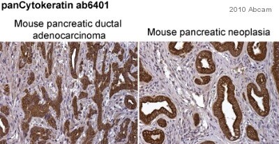
ab6401 staining pan Cytokeratin in mouse pancreatic ductal adenocarcinoma (left-hand panel) and mouse pancreatic neoplasia (right-hand panel) sections by immunohistochemistry (IHC-P – paraformaldehyde-fixed, paraffin-embedded sections). Tissue samples were fixed with paraformaldehyde and blocked with 10% serum for 1 hour at room temperature; heat mediated antigen retrieval was performed. The sample was incubated with primary antibody (1/250) at 4°C for 8 hours. A Biotin-conjugated Goat polyclonal (1/1000) was used as the secondary antibody.
This image is courtesy of an anonymous Abreview
See Abreview
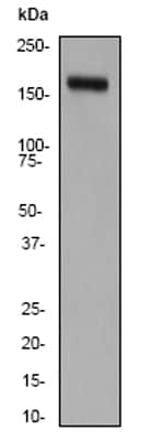
Western blot – pan Cytokeratin antibody [PCK-26] (ab6401)
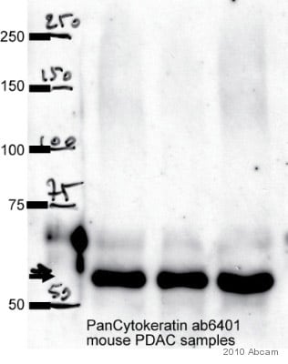
Observed band size : 55 kDa (why is the actual band size different from the predicted?)
All lanes: pan Cytokeratin antibody (ab6401) at 1/1000 dilution + whole cell lysate of Primary mouse pancreatic cancer cell lines (35 µg)
Secondary: An HRP-conjugated Sheep anti-mouse IgG polyclonal (1/5000) developed using the ECL technique
Performed under non-reducing conditions.
Blocking Step: 10% Milk for 1 hour at room temperature
This image is courtesy of an Abreview submitted by Pawel Mazur
See Abreview
Immunohistochemistry (Frozen sections) – pan Cytokeratin antibody [PCK-26] (ab6401)
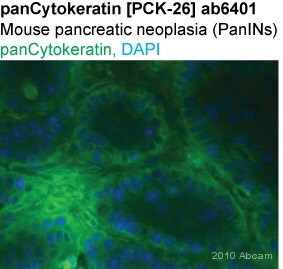
ab6401 staining pan Cytokeratin in Mouse pancreatic neoplasia tissue sections by Immunohistochemistry (IHC-Fr – frozen sections). Tissue was fixed with paraformaldehyde and blocked with 1% BSA for 1 hour at room temperature. Samples were incubated with primary antibody (1/250 in PBS) for 8 hours at 4°C. An Alexa Fluor®488-conjugated goat anti-mouse IgG polyclonal (1/1000) was used as the secondary antibody.
This image is courtesy of an anonymous Abreview
See Abreview
Immunocytochemistry/ Immunofluorescence – pan Cytokeratin antibody [PCK-26] (ab6401)
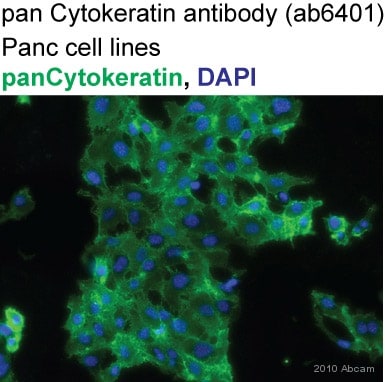
ab6401 staining pan Cytokeratin in Human Panc1 cells by ICC/IF (Immunocytochemistry/immunofluorescence). Cells were fixed with acetone/methanol (1:1) and blocked with 1% BSA for 1 hour at room temperature. Samples were incubated with primary antibody (1/300 in PBS) for 2 hours. An Alexa Fluor®488-conjugated DGoat anti-mouse IgG polyclonal (1/1000) was used as the secondary antibody. Nuclei were counterstained with DAPI.
This image is courtesy of an anonymous Abreview
See Abreview
References for Anti-pan Cytokeratin antibody [PCK-26] (ab6401)
Anti-pan Cytokeratin antibody [PCK-26] 鼠单克隆[PCK-26 详细产品信息可和选购


 Anti-Nitroguanosine polyclonal antibody


 (enlarge)
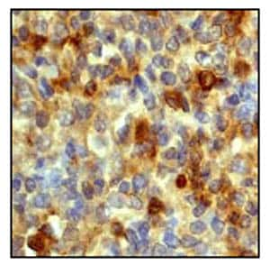 (enlarge)
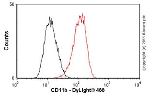 (enlarge)
