SOCS-2 Antibody (H-74)Santacruz抗体供应santacruz代理 SC-9022

SOCS-2 Antibody (H-74)Santacruz抗体供应

  • 产品型号:  santacruz代理 SC-9022
  • 简单描述
  • •rabbit polyclonal IgG, 200 µg/mlWB IP IF IHC(P) siRNA •epitope corresponding to amino acids 89-162 mapping within an internal region of SOCS-2 of human origin
详细介绍

此类产品还有其他抗体来源和种属反应详细产品信息请,谢谢!


ERK 2 (C-14) 抗体SC-154

ERK 2 (C-14) 抗体

  • 产品型号:  SC-154
  • 简单描述
  • ERK 2 (C-14) 抗体rabbit polyclonal IgG, 200 ?g/ml; also available as goat IgG, 200 ?g/ml, sc-154-G mouse, rat, human, avian, frog, zebrafish and Xenopus origin by WB, IP, IF, IHC(P), FCM and ELISA
详细介绍

ERK 2 (C-14) 抗体

ERK 2 Background Information
Mitogen-activated protein kinase (MAPK) signaling pathways involve two closely related MAP kinases, known as extracellular-signal-related kinase 1 (ERK 1, p44) and 2 (ERK 2, p42). Growth factors, steroid hormones, G protein-coupled receptor ligands, and neurotransmitters can initiate MAPK signaling pathways. Activation of ERK1 and ERK2 requires phosphorylation by upstream kinases such as MAP kinase kinase (MEK), MEK kinase and Raf-1. ERK1 and ERK2 phosphorylation can occur at specific tyrosine and threonine sites mapping within consensus motifs that include the Threonine-Glutamate-Tyrosine motif. ERK activation leads to dimerization with other ERKs and subsequent localization to the nucleus. Active ERK dimers phosphorylate serine and threonine residues on nuclear proteins and influence a host of responses that include proliferation, differentiation, transcription regulation and development. The human ERK2 gene maps to chromosome 22q11.21 and encodes a 360-amino acid protein.

  • rabbit polyclonal IgG, 200 µg/ml; also available as goat IgG, 200 µg/ml, sc-154-G
  • epitope mapping at the C-terminus of ERK 2 of rat origin
  • recommended for detection of ERK 2 p42 and, to a lesser extent, ERK 1 p44 of mouse, rat, human, avian, frog, zebrafish and Xenopus origin by WB, IP, IF, IHC(P), FCM and ELISA; also reactive with additional species, including equine, canine, bovine, porcine and avian
  • blocking peptide, sc-154 P
  • agarose conjugate for IP studies, sc-154 AC, 500 µg/0.25 ml agarose
  • fluorescein (sc-154 FITC) and rhodamine (sc-154 TRITC) conjugates for immunofluorescence, 200 µg/ml
  • phycoerythrin (sc-154 PE) conjugate for intracellular FCM, 100 tests
  • Alexa Fluor® 405 (sc-154 AF405), Alexa Fluor® 488 (sc-154 AF488) and Alexa Fluor® 647 (sc-154 AF647) conjugates for FCM or immunofluorescence; 100 µg/2 ml


ERK 2 (C-14) 抗体SC-154

ERK 2 (C-14): sc-154. Western blot analysis of ERK 2 expression in A-431 (A), HeLa (B), KNRK (C), NIH/3T3 (D), NRK (E) and DU 145 (F) whole cell lysates.

ERK 2 (C-14) 抗体SC-154

ERK 2 (C-14)-G: sc-154-G. Western blot analysis of ERK 1 and ERK 2 expression in A-431 (A), HeLa (B), KNRK (C) and NIH/3T3 (D) whole cell lysates.

ERK 2 (C-14) 抗体SC-154

ERK 2 (C-14): sc-154. Immunofluorescence staining of methanol-fixed HeLa cells (A) and immunoperoxidase staining of formalin-fixed, paraffin-embedded human breast carcinoma tissue (B) showing cytoplasmic localization.

ERK 2 (C-14) 抗体SC-154

Western blot analysis of ERK in 3611-RF (A,D), NIH/3T3 (B,E) and KNRK (C,F) whole cell lysates. Antibodies tested include ERK 1 (K-23)-G: sc-94-G (A-C) and ERK 2 (C-14)-G: sc-154-G (D-F).

ERK 2 (C-14) 抗体SC-154

ERK 2 (C-14) Alexa Fluor 488: sc-154 AF488. Intracellular FCM analysis of fixed and permeabilized HeLa cells. Black line histogram represents the isotype control, normal rabbit IgG: sc-45068.

ERK 2 (C-14) 抗体SC-154

ERK 2 (C-14) Alexa Fluor 647: sc-154 AF647. Intracellular FCM analysis of fixed and permeabilized HeLa cells. Black line histogram represents the isotype control, normal rabbit IgG: sc-24647.

ERK 2 (C-14) 抗体SC-154

ERK 2 (C-14): sc-154. Immunofluorescence staining of methanol-fixed HeLa cells showing cytoplasmic localization using indirect FITC (A) staining and direct Alexa Fluor 488 (B) staining.

ERK 2 (C-14) 抗体SC-154

ERK 2 (C-14): sc-154. Western blot analysis of ERK 1 expression in non-transfected 293T: sc-117752 (A), mouse ERK 1 transfected 293T: sc-126806 (B) and A-431 (C) whole cell lysates.

ERK 2 (C-14) 抗体SC-154

ERK 2 (C-14): sc-154. Western blot analysis of ERK 2 expression in rat hippocampus tissue extract.

ERK 2 (C-14) 抗体SC-154

ERK 2 (C-14)-G: sc-154-G. Western blot analysis of ERK 2 expression in rat hippocampus tissue extract.

ERK 2 (C-14) 抗体SC-154

ERK 2 (C-14) PE: sc-154 PE. Intracellular FCM analysis of fixed and permeabilized NIH/3T3 cells. Black line histogram represents the isotype control, normal rabbit IgG: sc-3871.

ERK 2 (C-14) 抗体SC-154

ERK 2 (C-14)-G: sc-154-G. Western blot analysis of ERK 2 expression in non-transfected 293T: sc-117752 (A), human ERK 2 transfected 293T: sc-177196 (B), A-431 (C) and HeLa (D) whole cell lysates.

ERK 2 (C-14) 抗体SC-154

ERK 1 (C-14): sc-154. Western blot analysis of ERK 1 expression in non-transfected: sc-117752 (A) and human ERK 1 transfected: sc-171765 (B) 293T whole cell lysates.


santacruz PCGF6 (E-15) 抗体*SC-160649

santacruz PCGF6 (E-15) 抗体*

  • 产品型号:  SC-160649
  • 简单描述
  • santacruz PCGF6 (E-15) 抗体*Epitope: Internal (h) Applications: WB, IF, ELISA Species: m, r, hIsotype: goat IgG
详细介绍

santacruz PCGF6 (E-15) 抗体*

BACKGROUND
Polycomb group (PcG) proteins form multiprotein complexes that regulate
expression patterns of developmental and cell proliferation genes. Several
members of the PcG contain ring finger domains and are identified as a subclass
of RING finger proteins. The RING-type zinc finger motif is present in a
number of viral and eukaryotic proteins and is made of a conserved cysteinerich
domain that is able to bind two zinc atoms. Proteins that contain the
RING-type zinc finger conserved domain are generally involved in the ubiquitination
pathway of protein degradation. PCGF6 (polycomb group ring finger
6), also known as MBLR or RNF134, is a 350 amino acid nuclear protein
that is ubiquitously expressed and contains one RING-type zinc finger. PCGF6
acts as a transcriptional repressor and regulates the level of histone
H3K4Me3 by activating SmcY histone demethylase. PCGF6 exists as two
alternatively spliced isoforms.
REFERENCES
1. Kanno, M., Hasegawa, M., Ishida, A., Isono, K. and Taniguchi, M. 1995.
Mel-18, a Polycomb group-related mammalian gene, encodes a transcriptional
negative regulator with tumor suppressive activity. EMBO J. 14:
5672-5678.
2. Satijn, D.P., Gunster, M.J., van der Vlag, J., Hamer, K.M., Schul, W.,
Alkema, M.J., Saurin, A.J., Freemont, P.S., van Driel, R. and Otte, A.P.
1997. RING1 is associated with the polycomb group protein complex and
acts as a transcriptional repressor. Mol. Cell. Biol. 17: 4105-4113.
3. Joazeiro, C.A. and Weissman, A.M. 2000. RING finger proteins: mediators
of ubiquitin ligase activity. Cell 102: 549-552.
4. Akasaka, T., Takahashi, N., Suzuki, M., Koseki, H., Bodmer, R. and Koga, H.
2002. MBLR, a new RING finger protein resembling mammalian Polycomb
gene products, is regulated by cell cycle-dependent phosphorylation. Genes
Cells 7: 835-850.
5. Tuckfield, A., Clouston, D.R., Wilanowski, T.M., Zhao, L.L., Cunningham,
J.M. and Jane, S.M. 2002. Binding of the RING polycomb proteins to specific
target genes in complex with the grainyhead-like family of developmental
transcription factors. Mol. Cell. Biol. 22: 1936-1946.
6. Online Mendelian Inheritance in Man, OMIM™. 2003. Johns Hopkins
University, Baltimore, MD. MIM Number: 607816. World Wide Web URL:
http://www.ncbi.nlm.nih.gov/omim/
7. Moore, R. and Boyd, L. 2004. Analysis of RING finger genes required for
embryogenesis in C. elegans. Genesis 38: 1-12.
8. Yang, Y., Lorick, K.L., Jensen, J.P. and Weissman, A.M. 2005. Expression
and evaluation of RING finger proteins. Meth. Enzymol. 398: 103-112.
9. Lee, M.G., Norman, J., Shilatifard, A. and Shiekhattar, R. 2007. Physical and
functional association of a trimethyl H3K4 demethylase and Ring6a/MBLR,
a polycomb-like protein. Cell 128: 877-887.
STORAGE
Store at 4° C, **DO NOT FREEZE**. Stable for one year from the date of
shipment. Non-hazardous. No MSDS required.
CHROMOSOMAL LOCATION
Genetic locus: PCGF6 (human) mapping to 10q24.33; Pcgf6 (mouse) mapping
to 19 C3.
SOURCE
PCGF6 (E-15) is an affinity purified goat polyclonal antibody raised against a
peptide mapping within an internal region of PCGF6 of human origin.
PRODUCT
Each vial contains 200 μg IgG in 1.0 ml of PBS with < 0.1% sodium azide
and 0.1% gelatin.
Blocking peptide available for competition studies, sc-160649 P, (100 μg
peptide in 0.5 ml PBS containing < 0.1% sodium azide and 0.2% BSA).
APPLICATIONS
PCGF6 (E-15) is recommended for detection of PCGF6 of mouse, rat and
human origin by Western Blotting (starting dilution 1:200, dilution range
1:100-1:1000), immunofluorescence (starting dilution 1:50, dilution range
1:50-1:500) and solid phase ELISA (starting dilution 1:30, dilution range
1:30-1:3000); non cross-reactive with PCGF1, PCGF3 or PCGF5.
Suitable for use as control antibody for PCGF6 siRNA (h): sc-90663, PCGF6
siRNA (m): sc-152109, PCGF6 shRNA Plasmid (h): sc-90663-SH, PCGF6
shRNA Plasmid (m): sc-152109-SH, PCGF6 shRNA (h) Lentiviral Particles:
sc-90663-V and PCGF6 shRNA (m) Lentiviral Particles: sc-152109-V.
Molecular Weight of PCGF6: 60 kDa.
RECOMMENDED SECONDARY REAGENTS
To ensure optimal results, the following support (secondary) reagents are
recommended: 1) Western Blotting: use donkey anti-goat IgG-HRP: sc-2020
(dilution range: 1:2000-1:100,000) or Cruz Marker™ compatible donkey
anti-goat IgG-HRP: sc-2033 (dilution range: 1:2000-1:5000), Cruz Marker™
Molecular Weight Standards: sc-2035, TBS Blotto A Blocking Reagent:
sc-2333 and Western Blotting Luminol Reagent: sc-2048. 2) Immunofluorescence:
use donkey anti-goat IgG-FITC: sc-2024 (dilution range: 1:100-
1:400) or donkey anti-goat IgG-TR: sc-2783 (dilution range: 1:100-1:400)
with UltraCruz™ Mounting Medium: sc-24941.
RESEARCH USE
For research use only, not for use in diagnostic procedures.
PROTOCOLS


高纯度有机溶剂及酸(SC级别)

高纯度有机溶剂及酸(SC级别)

  • 产品特性
  • 相关资料
  • Q&A
  • 参考文献

高纯度有机溶剂及酸(SC级别)高纯度有机溶剂及酸(SC级别)


  本产品适用于半导体行业,减少颗粒和金属杂质的电子级别高纯度试剂(SC级别)。

产品编号 产品名称 产品规格 产品等级 产品价格
018-23121 Acetone丙酮 1L SC
711-48001 Acetone丙酮 1L×8 SC
050-08001 Ethanol乙醇 1L SC
759-48001 Ethanol乙醇 1L×8 SC
133-16391 Methanol甲醇 1L SC
836-48001 Methanol甲醇 1L×8 SC
139-16871 N-Methyl-2-pyrrolidoneN-甲基-2-吡咯烷酮 1L SC
835-48211 N-Methyl-2-pyrrolidoneN-甲基-2-吡咯烷酮 1L×12 SC
089-09331 Hydrochloric Acid盐酸 1kg SC
785-48001 Hydrochloric Acid盐酸 1kg×12 SC
194-15761 Sulfuric Acid硫酸 1kg×12 SC
898-48001 Sulfuric Acid硫酸 1kg×12 SC

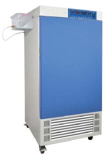
精品恒温恒湿培养箱LHS-300SC/LHS-150SCLHS-300SC/LHS-150SC

精品恒温恒湿培养箱LHS-300SC/LHS-150SC

简要描述:精品恒温恒湿培养箱LHS-300SC/LHS-150SC恒温恒湿箱有着精确的温度和湿度控制系统,它为产业研究、生物技术测试提供所需要的各种环境模拟条件,因此可广泛适用于药物、纺织、食品加工等无菌试验、稳定性检查以及工业产品的原料性能、产品包装、产品寿命等测试。

产品型号: LHS-300SC/LHS-150SC

所属分类:培养箱

详细说明:
品牌 其他品牌 价格区间 5千-1万
应用领域 医疗卫生,环保,化工,生物产业,农业

1、用途概述
恒温恒湿箱有着精确的温度和湿度控制系统,它为产业研究、生物技术测试提供所需要的各种环境模拟条件,因此可广泛适用于药物、纺织、食品加工等无菌试验、稳定性检查以及工业产品的原料性能、产品包装、产品寿命等测试。
精品恒温恒湿培养箱LHS-300SC/LHS-150SC
2、产品特点
n 人性化设计
⊕大屏幕液晶显示,多组数据一屏显示,菜单式操作界面,简单易懂,便于操作。
⊕致力于世界环保健康倡议,使用134 a,406 a无氟制冷剂,高效,节能,令环境更健康!
⊕采用镜面不锈钢内胆,四角半圆弧设计易清洁,箱内搁板间距可调。
⊕采用双层门结构,隔热性能好,观察箱内情况时不影响箱内温度。
⊕箱体左侧有一直径25mm/50mm的测试孔,便于实验操作与测量温度。(选配)
⊕可选箱门观察窗(选配、SC除外)
精品恒温恒湿培养箱LHS-300SC/LHS-150SC

精品恒温恒湿培养箱LHS-300SC/LHS-150SC
精品恒温恒湿培养箱LHS-300SC/LHS-150SCLHS-300SC/LHS-150SC
n 品质保证
⊕采用品牌压缩机,自主研发的压缩机降温系统,可有效延长压缩机使用寿命。
⊕采用JAKEL管流循环风机,配上*设计的风道结构,制造出良好的空气循环系统,有效保证箱内温度均匀性。
⊕采用湿度传感器,自主设计的内置水箱电热管加湿系统,有效的保证湿度偏差。
⊕PID控制方式,控温、控湿精确波动小,菜单式操作页面,简单易懂,便于操作。
⊕具有99个周期的程式,每周期分30段,每段99小时99分钟的循环步骤,几乎可以满足所有复杂的实验过程。
◆独立限温报警系统(选配)
⊕超过限制温度即自动中断运行,并声光报警提示操作者。保证实验安全运行不发生意外。
◆方便的数据处理(选配)
⊕具有USB或RS485接口,可连接打印机或计算机,能记录温度参数的变化状况。(选配)
3、可选配件
独立限温控制器
嵌入式打印机
RS485接口和软件
测试孔25mm/50mm
箱门观察窗(SC除外)
精品恒温恒湿培养箱LHS-300SC/LHS-150SC

4、技术参数

型号 LHS-100CH LHS-150SC LHS-250SC LHS-300SC
电源电压 AC220V  50HZ
控温范围 RT+10~85℃ 有加湿15~45℃ 无加湿10~45℃
恒温波动度 ±1℃ 高温±0.5℃  低温±1.5℃
温度分辨率 0.1℃
控湿范围 80~95%RH 60~85%RH
输入功率 1100W 620W 730W 850W
工作室尺寸
W×D×H(mm)
450×380×590 480×400×780 580×500×850 580×540×950
外形尺寸
W×D×H(mm)
575×605×1160 605×625×1350 705×725×1525 705×765×1640
公称容积 100L 150L 250L 300L
载物托架(标配) 2pcs 3pcs
定时范围 1~9999min