1) T. Akaike, S. Okamoto, T. Sawa, J. Yoshitake, F. Tamura, K. Ichimori, K. Miyazaki, K. Sasamoto and H. Maeda, "8-nitroguanosine formation in viral pneumonia and its implication for pathogenesis", Proc. Natl. Acad. Sci. USA, 2003, 100, 685.
2) J. Yoshitake, T. Akaike, T. Akuta, F. Tamura, T. Ogura, H. Esumi and H. Maeda, "Nitric oxide as an endogenous mutagen for Sendai virus without antiviral activity", J. Virol., 2004, 78, 8709.
3) T. Sawa, M. H. Zaki, T. Okamoto, T. Akuta, Y. Tokutomi, S. Kim-Mitsuyama, H. Ihara, A. Kobayashi, M. Yamamoto, S. Fujii, H. Arimoto and T. Akaike, "Protein S-guanylation by the biological signal 8-nitroguanosine 3',5'-cyclic monophosphate", Nat. Chem. Biol., 2007, 3, 727.
4) M. H. Zaki, S. Fujii, T. Okamoto, S. Islam, S. Khan, K. A. Ahmed, T. Sawa and T. Akaike, "Cytoprotective function of heme oxygenase 1 induced by a nitrated cyclic nucleotide formed during murine salmonellosis", J. Immunol., 2009, 182, 3746.
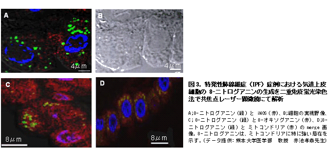
5) Y. Terasaki, T. Akuta, M. Terasaki, T. Sawa, T. Mori, T. Okamoto, M. Ozaki, M. Takeya and T. Akaike, "Guanine nitration in idiopathic pulmonary fibrosis and its implication for carcinogenesis", Am. J. Respir. Crit. Care. Med., 2006, 174, 665.
6) T. Sawa, M. Tatemichi, T. Akaike, A. Barbin and H. Ohshima, " Analysis of urinary 8-nitroguanine, a marker of nitrative nucleic acid damage, by high-performance liquid chromatography-electrochemical detection coupled with immunoaffinity purification: association with cigarette smoking", Free Radic. Biol. Med., 2006, 40, 711.
7) M. Feelisch, "Nitrated cyclic GMP as a new cellular signal", Nat. Chem. Biol., 2007, 3, 687.
8) K. A. Ahmed, T. Sawa and T. Akaike, "Protein cysteine S-guanylation and electrophilic signal transduction by endogeneous nitro-nucleotides", Amino Acids, 2011, 41(1), 123.
9) T. Sawa, H. Arimoto and T. Akaike, "Regulation of redox signaling involving chemical conjugation of protein thiols by nitric oxide and electrophiles", Bioconjugate Chem., 2010, 21(7), 1121


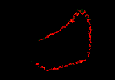
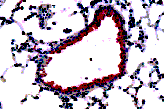
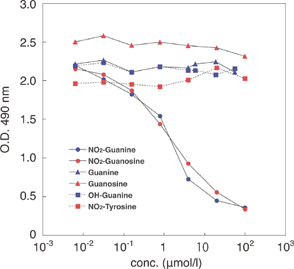
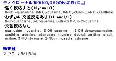
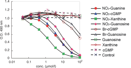
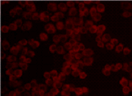
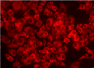
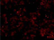
 Anti-Nitroguanosine polyclonal antibody