The SK7 (Leu-4) monoclonal antibody specifically binds to the epsilon chain of the CD3 antigen/T-cell antigen receptor (TCR) complex. This complex is composed of at least six proteins that range in molecular weight from 20 to 30 kDa. The antigen recognized by CD3 antibodies is noncovalently associated with either α/β or γ/δ TCR (70 to 90 kDa). The CD3 antigen is present on 61% to 85% of normal peripheral blood lymphocytes 60% to 85% of thymocytes and on Purkinje cells in cerebellum. The soluble form of this antibody has a mitogenic effect on most peripheral blood T lymphocytes, provided appropriate functional monocytes are present.
Aqueous buffered solution containing BSA and ≤0.09% sodium azide.
557851
FORMAT
Format
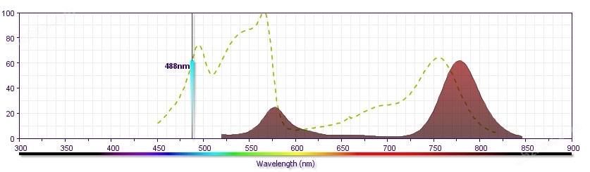
PE-Cy™7
Excitation Source
Blue 488 nm,Green 532 nm,Yellow/Green 561 nm
Excitation Max
496 nm
Emission Max
785 nm
PE-Cy™7 is a tandem fluorochrome that combines PE and a cyanine dye. PE-Cy7 conjugated reagents are as bright as PE conjugates. PE-Cy7 is particularly sensitive to photo-induced degradation, resulting in loss of fluorescence and changes in fluorescence spillover. Extreme caution must be taken to avoid light exposure and prolonged exposure to paraformaldehyde fixative. Fixed cells should be analyzed within 4 hours of fixation in paraformaldehyde or transferred to a paraformaldehyde-free buffer for overnight storage.
The RPA-T4 monoclonal antibody specifically binds to CD4, a 59 kDa single-chain transmembrane glycoprotein that is expressed on T-helper/inducer cell populations. CD4 is also expressed on thymocytes subsets and at lower levels on monocytes and macrophages. CD4 functions as a co-receptor in MHC class II-restricted antigen-induced T cell activation and as a receptor for human immunodeficiency viruses (HIV). This antibody binds to the D1 domain (CDR1 and CDR3 epitopes) of the CD4 antigen and reacts with approximay 80% of thymocytes and 45% of peripheral blood lymphocytes. RPA-T4 is capable of blocking HIV-1, gp120, and inhibits syncytium formation.
This antibody is routinely tested by flow cytometric analysis. Other applications were tested at BD Biosciences Pharmingen during antibody development or are reported in the literature.
FORMAT
Format
PE
Excitation Source
Blue 488 nm,Green 532 nm,Yellow/Green 561 nm
Excitation Max
496 nm
Emission Max
578 nm
R-phycoerythrin (PE) is an accessory photosynthetic pigment found in red algae. It exists in vitro as a 240-kDa protein with 23 phycoerythrobilin chromophores per molecule. This makes PE one of the brightest fluorochromes for flow cytometry applications, but its photobleaching properties make it unsuitable for fluorescence microscopy.
Other Formats →
APC
APC-Cy™7
APC-H7
APC-R700
Alexa Fluor® 488
Alexa Fluor® 647
Alexa Fluor® 700
BB515
BUV395
BV421
BV605
Biotin
FITC
NA/LE
PE-CF594
PE-Cy™5
PE-Cy™7
Pacific Blue™
PerCP-Cy™5.5
Purified
SUGGESTED COMPANION PRODUCTS
Cat No.
Description
Size
555749
PE Mouse IgG1, κ Isotype Control RUO
100 Tests
RESOURCES & TOOLS
Spectrum Viewer
Panel Designer
Download TDS
PREPARATION AND STORAGE
The monoclonal antibody was purified from tissue culture supernatant or ascites by affinity chromatography. The antibody was conjugated with R-PE under optimum conditions, and unconjugated antibody and free PE were removed. Store undiluted at 4°C and protected from prolonged exposure to light. Do not freeze.
PRODUCT NOTICES
This reagent has been pre-diluted for use at the recommended Volume per Test. We typically use 1 × 10^6 cells in a 100-µl experimental sample (a test).
Since applications vary, each investigator should titrate the reagent to obtain optimal results.
Please refer to www.bdbiosciences。。com/pharmingen/protocols for technical protocols.
For fluorochrome spectra and suitable instrument settings, please refer to our Multicolor Flow Cytometry web page at www.bdbiosciences。。com/colors.
Caution: Sodium azide yields highly toxic hydrazoic acid under acidic conditions. Dilute azide compounds in running water before discarding to avoid accumulation of potentially explosive deposits in plumbing.
Source of all serum proteins is from USDA inspected abattoirs located in the United States.
BD human CD16 PE-CY7 MAB抗体宿主:mouse反应物种:human规格:100T应用:流逝
详细介绍
BD human CD16 PE-CY7 MAB抗体
英文名:PE-Cy™7 Mouse Anti-Human CD16
克隆号:Clone 3G8 (RUO)
BD human CD16 PE-CY7 MAB抗体
介绍:
Brand
BD Pharmingen™
Vol. Per Test
5 µl
Isotype
Mouse BALB/c x DBA/2, also known as CD2F1 or CDF1 IgG1, κ
Reactivity
Human (QC Testing) Rhesus, Cynomolgus, Baboon (Reported)
Application
Flow cytometry (Routinely Tested)
Immunogen
Human polymorphonuclear leukocytes
Workshop No.
IV N409
Entrez Gene ID
2214 2215
Storage Buffer
Aqueous buffered solution containing BSA and ≤0.09% sodium azide.
Regulatory Status
RUO
规格
100 Tests 25 Tests
货号
557744
Product Details
References
DESCRIPTION
The 3G8 monoclonal antibody specifically binds to the 50-65 kDa transmembrane form of the IgG Fc Receptor (FcγRIII), a human NK cell-associated antigen. CD16 is expressed on NK cells as well as macrophages and granulocytes. Reports indicate that CD16 plays a role in signal transduction and NK cell activation. The 3G8 antibody blocks the binding of soluble immune complexes to granulocytes. The 3G8 antibody is reported (Vossebeld et al., 1997) to increase intracellular calcium levels in human neutrophils by interacting with bothFcγRIIa and FcγRIIIb molecules. This antibody has also been reported to induce homotypic neutrophil aggregation.
FORMAT
Format
PE-Cy™7
Excitation Source
Blue 488 nm,Green 532 nm,Yellow/Green 561 nm
Excitation Max
496 nm
Emission Max
785 nm
PE-Cy™7 is a tandem fluorochrome that combines PE and a cyanine dye. PE-Cy7 conjugated reagents are as bright as PE conjugates. PE-Cy7 is particularly sensitive to photo-induced degradation, resulting in loss of fluorescence and changes in fluorescence spillover. Extreme caution must be taken to avoid light exposure and prolonged exposure to paraformaldehyde fixative. Fixed cells should be analyzed within 4 hours of fixation in paraformaldehyde or transferred to a paraformaldehyde-free buffer for overnight storage.
Other Formats →
APC
APC-Cy™7
APC-H7
Alexa Fluor® 647
Alexa Fluor® 700
BUV395
BUV496
BUV737
BV421
BV510
BV605
BV650
BV711
BV786
Biotin
FITC
NA/LE
PE
PE-CF594
PE-Cy™5
SUGGESTED COMPANION PRODUCTS
Cat No.
Description
Size
557872
PE-Cy™7 Mouse IgG1 κ Isotype Control RUO
100 Tests
RESOURCES & TOOLS
Spectrum Viewer
Panel Designer
Download TDS
PREPARATION AND STORAGE
The monoclonal antibody was purified from tissue culture supernatant or ascites by affinity chromatography. The antibody was conjugated with PE-Cy7 under optimum conditions, and unconjugated antibody and free PE-Cy7 were removed. Store undiluted at 4°C and protected from prolonged exposure to light. Do not freeze.
Casciola-Rosen L, Rosen A, Petri M, Schlissel M. Surface blebs on apoptotic cells are sites of enhanced procoagulant activity: implications for coagulation events
and antigenic spread in systemic lupus erythematosus. Proc Natl Acad Sci U S A. 1996; 93(4):1624-1629. (Biology)
Homburg CH, de Haas M, von dem Borne AE, Verhoeven AJ, Reuingsperger CP, Roos D. Human neutrophils lose their surface Fc gamma RIII and acquire
Annexin V binding sites during apoptosis in vitro. Blood. 1995; 85(2):532-540. (Biology)
Koopman G, Reuingsperger CP, Kuijten GA, Keehnen RM, Pals ST, van Oers MH. Annexin V for flow cytometric detection of phosphatidylserine expression on
B cells undergoing apoptosis. Blood. 1994; 84(5):1415-1420. (Biology)
Martin SJ, Reuingsperger CP, McGahon AJ, et al. Early redistribution of plasma membrane phosphatidylserine is a general feature of apoptosis regardless of
the initiating stimulus: inhibition by overexpression of Bcl-2 and Abl. J Exp Med. 1995; 182(5):1545-1556. (Biology)
Raynal P, Pollard HB. Annexins: the problem of assessing the biological role for a gene family of multifunctional calcium- and phospholipid-binding proteins.
van Engeland M, Ramaekers FC, Schutte B, Reuingsperger CP. A novel assay to measure loss of plasma membrane asymmetry during apoptosis of adherent
cells in culture. Cytometry. 1996; 24(2):131-139. (Biology)
Vermes I, Haanen C, Steffens-Nakken H, Reuingsperger C. A novel assay for apoptosis. Flow cytometric detection of phosphatidylserine expression on early
apoptotic cells using fluorescein labelled Annexin V. J Immunol Methods. 1995; 184(1):39-51. (Biology)
BD细胞凋亡试剂盒(PE和7-ADD标记)PE Annexin V Apoptosis Detect
产品型号: 559763
简单描述
BD细胞凋亡试剂盒(PE和7-ADD标记)PE Annexin V Apoptosis Detection Kit I 100TESTNameAnnexin V : PE Apoptosis Detection Kit IContentsAnnexin V-PE, 7-AAD, and Annexin V Binding BufferSize100 TestsRegulatory Stat
详细介绍
BD细胞凋亡试剂盒(PE和7-ADD标记)PE Annexin V Apoptosis Detection Kit I
Technical Data Sheet
PE Annexin V Apoptosis Detection Kit I
Product Information
Material Number: 559763
Component: 51-66121E
Description: 10X Annexin V Binding Buffer
Size: 50 ml (1 ea)
Storage Buffer: Aqueous buffered solution containing no preservative.
Casciola-Rosen L, Rosen A, Petri M, Schlissel M. Surface blebs on apoptotic cells are sites of enhanced procoagulant activity: implications for coagulation events
and antigenic spread in systemic lupus erythematosus. Proc Natl Acad Sci U S A. 1996; 93(4):1624-1629. (Biology)
Homburg CH, de Haas M, von dem Borne AE, Verhoeven AJ, Reuingsperger CP, Roos D. Human neutrophils lose their surface Fc gamma RIII and acquire
Annexin V binding sites during apoptosis in vitro. Blood. 1995; 85(2):532-540. (Biology)
Koopman G, Reuingsperger CP, Kuijten GA, Keehnen RM, Pals ST, van Oers MH. Annexin V for flow cytometric detection of phosphatidylserine expression on
B cells undergoing apoptosis. Blood. 1994; 84(5):1415-1420. (Biology)
Martin SJ, Reuingsperger CP, McGahon AJ, et al. Early redistribution of plasma membrane phosphatidylserine is a general feature of apoptosis regardless of
the initiating stimulus: inhibition by overexpression of Bcl-2 and Abl. J Exp Med. 1995; 182(5):1545-1556. (Biology)
Raynal P, Pollard HB. Annexins: the problem of assessing the biological role for a gene family of multifunctional calcium- and phospholipid-binding proteins.
van Engeland M, Ramaekers FC, Schutte B, Reuingsperger CP. A novel assay to measure loss of plasma membrane asymmetry during apoptosis of adherent
cells in culture. Cytometry. 1996; 24(2):131-139. (Biology)
Vermes I, Haanen C, Steffens-Nakken H, Reuingsperger C. A novel assay for apoptosis. Flow cytometric detection of phosphatidylserine expression on early
apoptotic cells using fluorescein labelled Annexin V. J Immunol Methods. 1995; 184(1):39-51. (Biology)
Description: The BM8 monoclonal antibody reacts with mouse F4/80 antigen, an approximay 125 kDa transmembrane protein. The F4/80 antigen is expressed by a majority of mature macrophages and is the best marker for this population of cells. However, other cell types such as Langerhans cells and liver Kupffer cells have been reported to express this antigen. Expression of F4/80 commences during early myeloid development and is upregulated on all BM cells stimulated in vitro with M-CSF. It has been shown that some cytokines downregulate the expression of F4/80 resulting in lack of F4/80 antigen on a subpopulation of macrophages, especially in the lymphoid microenvironment in vivo